The Anatomy Scan: What Happens at Your 20-Week Ultrasound
A complete guide to the mid-pregnancy anatomy scan — what it examines, what it can and cannot detect, and how to prepare for what you might hear.
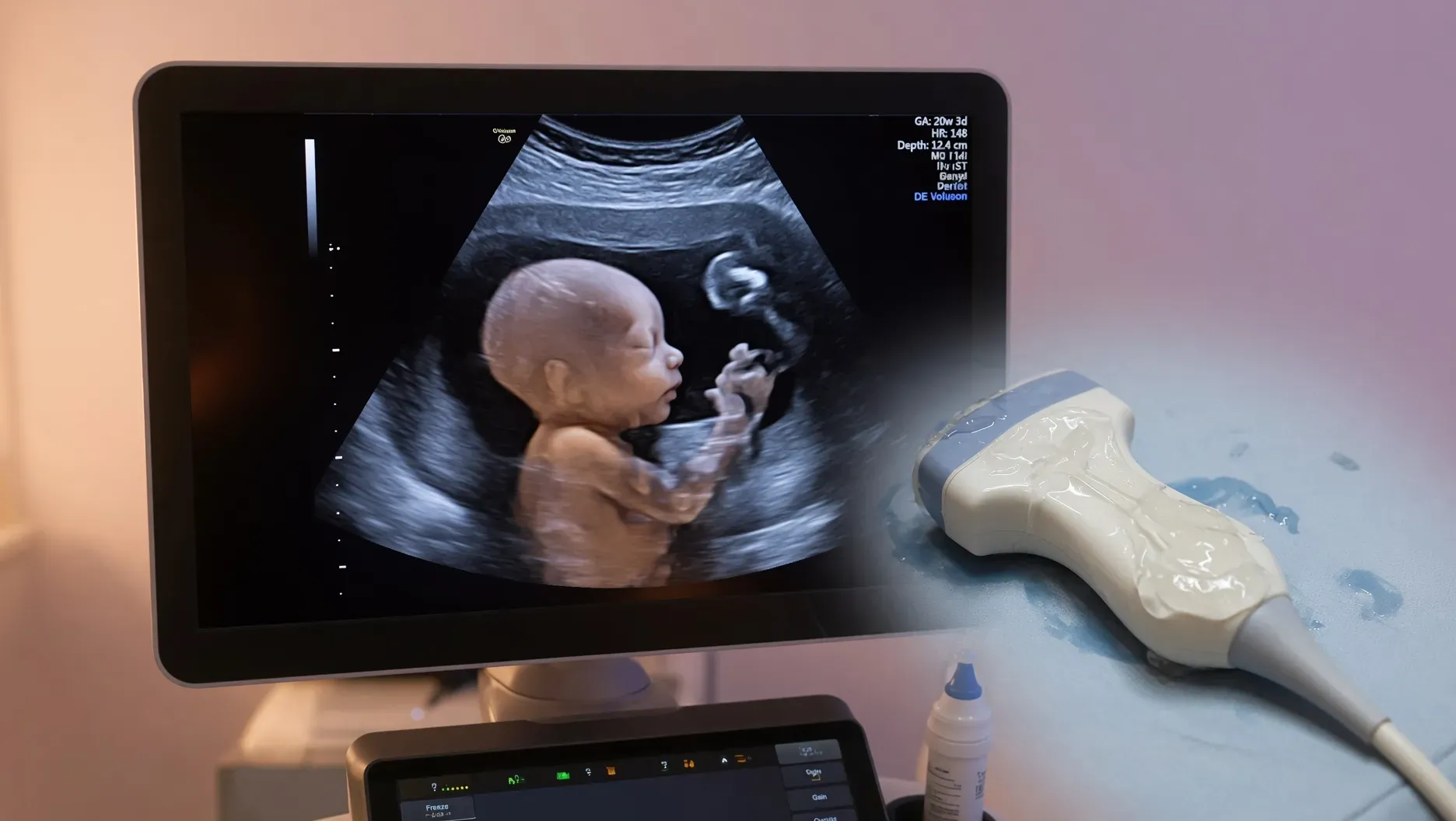
The mid-pregnancy ultrasound — commonly called the anatomy scan, anomaly scan, or level II ultrasound — is the most detailed examination of your baby that will happen before birth. It is the scan that most parents look forward to with a specific mix of excitement and quiet anxiety: the moment when the pregnancy becomes visually, undeniably real, and when a great deal of information about how the baby is developing is gathered in a single appointment.
Understanding what this scan is actually examining, what the sonographer is looking for, what it can and cannot detect, and how to prepare for the appointment — including for the possibility of findings that require follow-up — allows you to enter this appointment informed rather than uncertain.
When the anatomy scan happens
The anatomy scan is typically performed between eighteen and twenty-two weeks of pregnancy, with twenty to twenty-two weeks considered optimal. At this point in pregnancy:
- The baby is large enough that individual structures are clearly visible on ultrasound
- The baby is small enough that all structures fit within a single image and the full anatomy can be surveyed systematically
- If abnormalities are identified, there is still time for further investigations, specialist consultations, and, where relevant, decisions to be made before viability becomes a complex factor
In India, the scan is commonly referred to as the mid-trimester anomaly scan, the level II scan, or simply the twenty-week scan. It is typically performed at a radiology centre or maternity hospital equipped with a diagnostic ultrasound machine capable of detailed imaging.
What the scan examines
The anatomy scan is a systematic survey of your baby’s physical structures. A trained sonographer moves the probe methodically through a checklist of structures, measuring and documenting each one. The examination typically takes thirty to sixty minutes, though this varies depending on the baby’s position, movement, and cooperation.
The baby’s head and brain:
- Head circumference and shape
- The cerebral hemispheres and ventricles (fluid-filled spaces in the brain)
- The choroid plexus (structures within the ventricles that produce cerebrospinal fluid)
- The cerebellum (the structure at the back of the brain involved in coordination and balance)
- The nuchal fold (thickness at the back of the neck, a soft marker for chromosomal conditions)
- The face — profile, lips, and nasal bone
The spine:
- Viewed along its length and in cross-section
- Looking for open defects such as spina bifida, in which the spinal column fails to close properly
The heart:
- Four-chamber view, confirming the presence and relative size of all four heart chambers
- Outflow tracts — the major vessels leaving the heart
- Rhythm and rate
- Position within the chest
Cardiac abnormalities are among the most common birth defects and can be challenging to visualise fully on ultrasound. Some facilities perform a more detailed cardiac assessment (fetal echocardiography) separately, particularly for high-risk pregnancies.
The abdominal organs:
- Stomach — should be visible as a fluid-filled bubble, confirming the baby is swallowing amniotic fluid
- Kidneys — both kidneys should be present and of appropriate size and appearance
- Bladder — should be visible as a fluid-filled structure
- Abdominal wall — confirming closure; open defects such as gastroschisis and omphalocele are looked for here
The limbs:
- All four limbs should be present
- Long bone lengths (femur, humerus) are measured
- Hands and feet are visualised, though detailed finger counting is not always possible
Growth measurements:
- Head circumference
- Abdominal circumference
- Femur (thigh bone) length
- Biparietal diameter (width across the head)
These measurements are combined to estimate the baby’s weight and are plotted on a growth chart to assess whether growth is appropriate for gestational age.
The placenta:
- Location — the placenta’s position relative to the cervix is specifically assessed
- Placenta praevia — when the placenta lies low and covers or is very close to the cervical opening — is identified here. Low-lying placenta at twenty weeks is relatively common and often resolves by later in pregnancy as the uterus grows; however, persistent praevia at thirty-two to thirty-six weeks is clinically significant and affects delivery planning.
Amniotic fluid:
- The amniotic fluid index (AFI) is measured — too little (oligohydramnios) or too much (polyhydramnios) fluid can both indicate issues with fetal wellbeing
The cervix:
- Sometimes assessed separately, either transabdominally or transvaginally, for length — a short cervix is a risk factor for preterm birth
What the scan can and cannot detect
This is important to understand before the appointment, because the anatomy scan is a screening tool, not a diagnostic test that catches everything.
What it can detect: Major structural abnormalities in the organs and systems it examines. Neural tube defects, major heart defects, abdominal wall defects, severe limb abnormalities, abnormalities of the kidneys and urinary tract, and significant brain structural problems are among those detectable when present and visible.
What it cannot reliably detect: Small heart defects (including many ventricular septal defects), chromosomal conditions without structural features visible on ultrasound (including Down syndrome, though structural soft markers may be present), conditions that develop after twenty weeks, and abnormalities too subtle to be resolved by current ultrasound technology. It also cannot rule out genetic conditions, developmental delay, autism, or behavioural conditions — none of these have structural ultrasound features detectable at this stage.
Detection rates vary by condition and facility: The detection rate for major abnormalities ranges from forty to ninety percent depending on the condition, the quality of the equipment, the experience of the sonographer, and factors outside anyone’s control — primarily the baby’s position. A baby lying in an unfavourable position may obscure specific structures.
The anatomy scan is the most thorough available examination before birth, but a normal anatomy scan does not guarantee a baby without any condition. This is not a limitation of the scan — it is the honest reality of what ultrasound can and cannot visualise.
A note on sex determination in India
In India, prenatal sex determination — revealing the sex of the baby through ultrasound — is illegal under the Pre-Conception and Pre-Natal Diagnostic Techniques (PCPNDT) Act of 1994, due to the documented misuse of this information leading to sex-selective abortions and a severely skewed sex ratio.
Sonographers are legally prohibited from revealing or indicating the sex of the baby during any prenatal scan. This applies to the anatomy scan. It is not a hospital policy or individual choice — it is the law, and the consequences for violations are serious.
What happens if something is found
Finding that the scan has identified something concerning is one of the possibilities that parents approach this appointment with quiet anxiety about. Understanding what that process looks like — calmly and in advance — helps.
If the sonographer sees something that requires follow-up, a few things typically happen:
Referral for repeat or detailed scan: Some findings require re-examination at a later date (because the baby’s position prevented full assessment) or at a specialised centre with higher-resolution equipment or a sonographer with specific fetal medicine expertise.
Referral to a fetal medicine specialist: A maternal-fetal medicine specialist (perinatologist) reviews complex or concerning findings, interprets them in clinical context, and advises on next steps.
Further investigations: Depending on the finding, amniocentesis (sampling of amniotic fluid to test fetal chromosomes) or other investigations may be offered to provide diagnostic information. These are offered, not required — the decision is yours.
Counselling: A finding at the anatomy scan should be followed by detailed, honest counselling from your provider about what has been found, what it means in terms of probability and severity, what further information is available, and what the options are.
Not every finding at an anatomy scan is a confirmed problem. Some findings are incidental soft markers — features that are statistically more common in certain chromosomal conditions but are also present in many chromosomes-normal pregnancies. These require contextualisation rather than alarm. The conversation with your provider about what a specific finding means for your specific situation is the important one, not a general internet search.
How to prepare for the scan
Practical preparation:
- Most anatomy scans are performed transabdominally (the probe on your abdomen) and do not require a full bladder, though some facilities request one for the initial survey
- Wear clothing that allows easy access to the abdomen
- The appointment may take thirty to sixty minutes; plan accordingly
- You may be asked to walk around between attempts if the baby is not in a good position
Emotional preparation:
- Bring your partner or a support person if your facility allows and if you would like to
- Understand that the sonographer may be quiet and focused for long periods — they are working through a systematic checklist, and silence does not indicate concern
- Know before the appointment that the sonographer may not be able to answer detailed questions about findings — their role is to image and document; interpretation is done by the radiologist and your obstetrician
- The report goes to your provider, who will discuss it with you either immediately or at your next appointment
If you are anxious: This is normal. Most anatomy scans are normal. The scan exists precisely to catch problems early when they can be most effectively managed — which means that the value of going is the same whether the result is reassuring or whether it identifies something that needs follow-up.
The honest message
The anatomy scan is one of the most significant appointments of pregnancy. It takes a systematic, careful look at how your baby is developing. For the great majority of women, it is reassuring — and the relief of a normal result is real and earned.
For some women, it identifies something that requires further attention. That further attention, when needed, is exactly what the scan is designed to enable — earlier knowledge, more time to prepare, and access to specialist care. A finding at the anatomy scan is not the end of a story. It is, more often, the beginning of a more closely supported one.
Go to the scan. Know what it is examining. Let the sonographer do their work. And hear the results from your provider with the understanding that, whatever they are, you will have support in navigating what comes next.
This article is for general educational purposes only and does not replace personalised medical advice. Always consult your doctor, midwife, or a qualified healthcare professional about the results of any prenatal scan and what they mean for your pregnancy.